


Technique developed in mice could aid detection of cancer in dense breasts
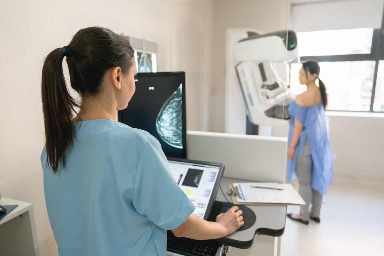
A two-pronged approach to imaging breast density in mice, developed by researchers at Georgetown Lombardi Comprehensive Cancer Center, has resulted in better detection of changes in breast tissue, including spotting early signs of cancer. The researchers hope that this approach will be translated from mice and improve breast imaging for people; it may also help with prognosis of disease as density can be linked to specific patterns of mammary gland growth, including signs of cancer development.
The findings appeared Sept. 14, 2022 in the American Journal of Pathology.
"Having a means to accurately assess mammary gland density in mice, just as is done clinically for women using mammograms, is an important research advance," says Priscilla A. Furth, MD, professor of oncology and medicine at Georgetown Lombardi and corresponding author of the study. "This method has the benefit of being applicable across all ages of mice and mammary gland shapes, unlike some methods used in earlier studies."
An innovative analytic computer program, developed by Georgetown alum Brendan Rooney while working as an undergraduate in Furth's lab, allowed for sorting of mammary gland tissue to one of two imaging assessments. Rooney initially looked at younger mouse glands and found that a program that removed background "noise" in those images helped boost detection of abnormalities in what are typically rounder, more lobular tissues.
Read more...
Quick Links


Powered by: 